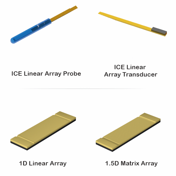
ICE Intracardiac Echocardiogram
ICE ultrasound probe, 2D-ICE probe, 4D-ICE probe. . .
ICE (Intracardiac Echocardiography) is an emerging technology that involves implanting miniature transducers at the tip of a catheter which is then guided through peripheral vessels to the heart’s interior. The transducers emit sound waves to produce real-time high-quality imaging and/or hemodynamic measurements of the heart and adjacent tissues.
ICE is a highly advanced technology that integrates state-of-the-art ultrasound and image processing, and represents one of the most challenging areas in the field of intravascular ultrasound today. ICE allows for direct visualization of cardiac structures which facilitates understanding of the anatomical relationships between different parts of the heart. It provides real-time imaging, complication monitoring, good tolerability, and can be performed through the femoral vein without the need for general anesthesia or deep sedation. Consequently, it has become an important adjunctive tool in cardiac interventional procedures and is often referred to as the “gold standard” for interventional cardiologists.

“With the increasing popularity and complexity of cardiac intervention procedures, the demand for intraoperative imaging is becoming higher. Intracardiac echocardiography (ICE) is well suited for cardiac intervention operations due to its real-time imaging, real-time monitoring of intraoperative complications, and good tolerability, among other advantages. It is particularly useful as it does not involve X-rays, allowing repetitive operations, full-range visualization, precise display of local anatomical structures and cardiac blood flow signals and velocity. As a result, ICE is increasingly used in various types of cardiac intervention procedures.
– “Consensus on Intracardiac Echocardiography in China”
Chinese Journal of Cardiac Pacing and Electrophysiology”


